Ophthalmology is a dynamically developing field of medicine concerning the organ of sight – the eyes. It covers the prevention, diagnosis, and treatment of eye diseases, which increasingly affect our patients.
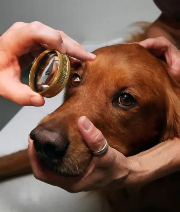
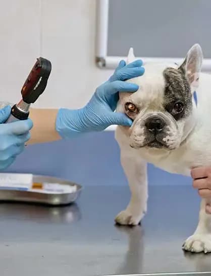
During an ophthalmic examination, the following are assessed, among others: the quantity and quality of the tear film produced, the structure of the eyeballs and the structures surrounding them, the presence of ectopic and/or distichiatic eyelashes, the structure of the eyelids, the intraocular pressure, the transparency of the optic media, the presence of corneal ulcers, and the reflexes.
Dog and cat owners most often come to a veterinary ophthalmologist in connection with the following problems:
Conjunctivitis
Conjunctivitis manifests as pain and/or itching, squinting of the eye, redness of the conjunctiva, and increased discharge from the conjunctival sacs. It may affect one or both eyes and be caused by an allergy, irritation, or infection, or it may have no known cause. If your veterinarian suspects conjunctivitis in your pet, they will carry out examinations aimed at ruling out concurrent diseases and selecting the appropriate treatment.
Corneal ulcers/ulceration
Corneal ulcers/ulceration are, to put it simply, a break in the continuity of the cornea, that is, the outermost layer of the eyeball. They are most often the result of mechanical trauma to the eye and manifest as severe pain, squinting of the eye, malaise, redness of the eye, and discharge.
If your dog or cat shows the above symptoms, seek a consultation without delay – corneal ulceration requires intensive and promptly implemented treatment. The veterinarian will perform a test with a dye – fluorescein – which makes it possible to detect damage to the surface of the eye.
"Tear stains" under the eyes
Rusty or brown "tear stains" with an unpleasant smell under the eyes are a common ailment of our pets. Contrary to appearances, they are not only a cosmetic problem but also a health one. The prolonged escape of tears onto the fur and skin under the eyes can lead to secondary bacterial and fungal infections, as well as to inflammation and maceration of the skin. It is the proliferation of Malassezia spp. fungi that is the cause of the unpleasant, "yeasty" smell of the stains. These fungi are very fond of warm and moist conditions, which is why they choose the tear-soaked area under the eyes as their habitat.
So what can be done to get rid of this problem? First, together with your veterinarian, you need to determine the cause of the excessive tearing. It may be caused by a food or environmental allergy, an infection, conjunctivitis, or an abnormal structure of the eye. When tear stains under the eyes occur, it is extremely important to book your dog in with a veterinary ophthalmologist in order to examine the whole eye, including its structure. Increasingly, the cause is the presence of an eyelid defect – entropion – or the presence of additional, distichiatic or ectopic eyelashes (we discuss these problems below). Examining the organ of sight makes it possible to select the appropriate treatment and often to eliminate the problem entirely.
Entropion
Entropion is an eyelid structural defect in which the eyelids roll inward toward the eyeball, irritating its surface and often preventing the drainage of tears through the nasolacrimal ducts. A similar defect is ectropion, in which the eyelids roll outward. The presence of this defect secondarily leads to the formation of tear stains under the eyes, conjunctivitis, excessive tearing, infections, and corneal damage. Dogs suffering from entropion may experience discomfort. This problem is treated surgically. Often this surgical procedure makes it possible to eliminate or limit the pet's secondary problems, such as the formation of tear stains under the eyes, recurrent conjunctivitis, or corneal damage.
Distichiatic/ectopic eyelashes
Distichiatic eyelashes – these are additional, supernumerary eyelashes located on the eyelid margin. They may be directed toward the eye and irritate the surface of the cornea.
Ectopic eyelashes – these are additional eyelashes located in an abnormal place, growing out of the conjunctiva of the eye and irritating the cornea.
They may contribute to the formation of tear stains under the eyes, conjunctivitis, excessive tearing, infections, corneal damage, and the animal feeling discomfort. Both distichiatic and ectopic eyelashes are removed under general anaesthesia. Often this procedure makes it possible to limit or eliminate the pet's secondary problems, such as the formation of tear stains under the eyes, recurrent conjunctivitis, or corneal damage.
Cataract
A cataract is a clouding of the eye's lens, which can lead to partial or total blindness. It may involve the entire lens or only a part of it. In an advanced stage, a cataract is visible to the owner with the naked eye, giving the effect of a cloudy, "milky" eye. This is a condition that we most associate with older animals (the so-called senile cataract); however, it can also occur in younger individuals.
We distinguish four main types of cataract:
- senile – occurs in older animals as a result of the ageing of the tissues,
- congenital – this is a cataract that the animal is born with. It may, but does not have to, be hereditary,
- acquired – arises during the animal's life, e.g. as a result of an infection, injury, the use of certain medications, radiotherapy, or improper nutrition,
- diabetic – develops as a complication of diabetes.
Ophthalmic examination of animals intended for breeding
Owing to the existence of hereditary diseases of the organ of sight, it is extremely important to carry out an ophthalmic examination of purebred dogs and cats intended for breeding. This is particularly important in breeds predisposed to eye diseases. Examples of such breeds and diseases are:
- multifocal retinal dysplasia (MRD) in, among others, Cavalier King Charles Spaniels, Golden Retrievers, and Rottweilers;
- congenital cataract in, among others, Miniature Schnauzers, the Old English Sheepdog, and the West Highland White Terrier; Collie Eye Anomaly (CEA) in Border Collies, Rough Collies, and Shetland Sheepdogs;
- progressive retinal atrophy (PRA) in, among others, Irish Setters and the Cardigan Welsh Corgi.
Depending on the disease, in order to rule out the occurrence of hereditary eye diseases in the offspring, it is necessary to carry out an ophthalmic examination and/or genetic tests on the future parents.
We invite you to book a visit at our clinic and to ask any questions concerning your animal's eye health. We are here to help!
DVM Angelika Pietrzkiewicz