Kidney tumours in dogs are among the less commonly diagnosed, yet remarkably instructive, oncological conditions – precisely because they so often produce no symptoms for a long time. The story of Benio, a Cavalier King Charles Spaniel whose owner regularly brought him in for follow-up ultrasound scans, is a perfect illustration: his kidney tumour was found completely by chance, in a dog who felt great, ate with a healthy appetite, and showed no sign of illness whatsoever. This article is both a detailed account of Benio's case – including the test results and the course of treatment, exactly as they were recorded in his medical documentation – and an extensive guide to kidney tumours in dogs: why they're so often missed, how they're diagnosed, what surgical treatment involves, and why regular check-ups, even in animals that seem perfectly healthy, matter so much. We're describing this story step by step, the way it appears in Benio's medical records – including the precise ultrasound and histopathology findings – because this level of detail best illustrates what the journey from an incidental finding on a scan to a full diagnosis and recovery actually looks like in practice.
Benio's Story: An Incidental Discovery of a Kidney Tumour
Referral and History
Benio, a male dog of the Cavalier King Charles Spaniel breed, Blenheim colour, 4.5 years old, came to our clinic with a diagnosis of a tumour in his left kidney. The tumour was discovered during a routine ultrasound (USG) check-up at the "Kalinowa" clinic in Łódź, where Benio lives permanently. His owner had been prompted to schedule regular six-monthly check-ups by Benio's earlier medical history – a year before, he had gone through a bout of bladder inflammation, and at that time a urinalysis had detected ammonium magnesium phosphate crystals. Crucially, at the time he came to us, Benio showed no symptoms of illness whatsoever. He was in good condition, had a good appetite, and all his physiological functions were normal.

This part of Benio's story is one of the most important lessons the whole case offers: had it not been for the regular, routine check-ups scheduled after his earlier urinary episode, his kidney tumour would most likely have gone undetected for many more months, possibly even years.
Clinical Examination and Additional Tests
The clinical examination carried out at our clinic revealed no abnormalities – Benio was in good condition, had a good appetite, and all his physiological functions remained normal.
Additional tests – blood and urine analysis – showed no extreme deviations from normal. The only exception was a slightly elevated white blood cell count. That's a telling detail in itself: it shows just how unremarkable general test results can remain even in the presence of a genuine, several-centimetre tumour in one kidney – especially when the other kidney is working properly.
Ultrasound Findings
The result of the ultrasound (USG) examination was key to what followed. The liver was of normal size, with a normal parenchymal structure and no focal lesions. The gallbladder had no stones, and the bile ducts weren't dilated. The spleen wasn't enlarged, with no changes in the parenchyma.
The left kidney was enlarged at the lower pole, and in the central part of the kidney a solid lesion approximately 40 mm in diameter was visualised, with minor cystic remodelling suggestive of neoplastic growth. No inflammatory or infiltrative reaction to the surrounding structures was found around the lesion. The right kidney wasn't enlarged; its parenchyma was mildly remodelled, the renal pelvis wasn't dilated, and there were no stones.
The adrenal glands weren't enlarged, and the pancreas was unchanged. The bladder showed no structural changes and no visible stones. The prostate gland measured 33 x 32 mm, with a normal parenchymal structure and no cysts. No fluid was found in the abdominal cavity, and the lymph nodes weren't enlarged.
Surgery – Left Nephrectomy
On 11 January 2012, Benio was operated on. A left nephrectomy was performed – the surgical removal of the entire diseased kidney along with the tumour. After the procedure, Benio went home; photos from that time show him already resting on his blanket.
Histopathology Result
The removed kidney was sent for histopathological examination, which ultimately determined the diagnosis and the outlook going forward.
MACROSCOPIC FINDINGS: Kidney measuring 9x5x4 cm, with a smooth external surface. On cross-section, at one pole, a well-demarcated, cream-coloured tumour with a finely cystic cut surface, approximately 4 cm in diameter. The tumour does not reach the renal pelvis.
MICROSCOPIC FINDINGS: Carcinoma tubulare renis bene differentiatum (G-1). The tumour is composed of numerous tubular structures of varying size, with mild cytological atypia and occasional mitotic figures. The tumour is not encapsulated but is well demarcated from the remaining parenchyma; it does not reach the hilar structures and does not extend beyond the surface of the kidney. The hilar vessels contain no tumour metastases.
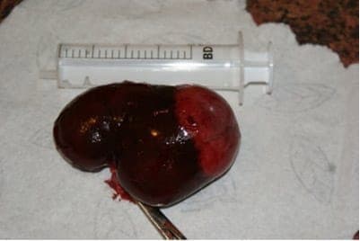
In practical terms, this result was good news: the tumour was well differentiated, had clear margins, hadn't invaded the renal hilar structures or blood vessels, and hadn't extended beyond the renal capsule. We explain exactly what these terms mean later in this article.
Case Summary
Benio is currently doing great; he remains under the care of the clinic in Łódź, where he lives. He swims, chases ducks, and is in excellent condition. His owner now pays much closer attention to keeping his weight in check. The only supplement Benio currently receives is omega-3 fatty acids from flaxseed oil, together with a rockrose (Cistus) preparation, given at a dose of one capsule a day. We hope this good state of health continues for a long time to come.
The purpose of describing this case is clear – we want to emphasise the importance of regular check-ups in animals: in young animals after an illness, and in older animals over 6–7 years of age, at least once a year.
This conclusion, stated directly by the team who treated Benio, is also the best bridge to the second part of this article – a broader look at kidney tumours in dogs: what they are, why they can be so insidious, and what their diagnosis and treatment involve.
Kidney Tumours in Dogs – What You Should Know
Primary kidney tumours – meaning ones that arise directly from the tissue of the kidney itself, as opposed to metastases from other organs – are rare in dogs. The kidneys are far more often a site where tumours that originated elsewhere spread to, rather than the site where a primary tumour develops. Among primary kidney tumours, the most commonly diagnosed type is renal carcinoma, often of tubular architecture – exactly the type diagnosed in Benio. Rarer types include nephroblastoma, which occurs more often in younger dogs, and various forms of sarcoma. Primary kidney tumours in dogs almost always affect a single kidney – as in Benio's case, where the lesion involved only the left kidney, while the right one remained functional and only slightly remodelled. Kidney tumours are usually diagnosed in middle-aged and older dogs, though – as Benio's case shows, since he was only 4.5 years old at the time of diagnosis – they can also appear in considerably younger animals. That's one reason vets are reluctant to "rule out" cancer purely on the basis of a patient's age, and why any unclear finding on imaging, regardless of the dog's age, warrants full investigation.
Why Kidney Tumours Go Unnoticed for So Long
Kidneys have an enormous functional reserve. As long as the disease affects only one of them, and the other is working properly, a dog's body can keep its blood and urine parameters normal for a very long time – exactly as happened with Benio, whose blood and urine tests showed only a slightly elevated white blood cell count, despite the presence of a several-centimetre tumour. That's why primary kidney tumours are so often discovered incidentally, during imaging carried out for a completely different reason – a check-up after a previous illness, a pre-surgical work-up, or a routine abdominal ultrasound in an older dog. Had Benio not had a bout of bladder inflammation a year earlier, and not been placed under regular ultrasound monitoring as a result, his kidney tumour would most likely have gone unnoticed until it started producing clear clinical signs – which usually means the disease has already reached a more advanced stage.
Symptoms That Can Appear at a More Advanced Stage
Benio had no symptoms at all – and that's actually the best-case scenario, because it meant the tumour was caught early. But as a kidney tumour grows, reaches a larger size, or begins to invade nearby structures, the following can appear:
- blood in the urine, sometimes visible to the naked eye, sometimes detected only in a laboratory test,
- a mass in the abdomen that can be felt on palpation,
- gradual weight loss,
- weakness and reduced exercise tolerance,
- a worsening or loss of appetite,
- in rarer cases, a fever of unclear origin.
Rarer still are so-called paraneoplastic syndromes – disturbances that are an indirect consequence of the tumour's presence, rather than a result of it directly invading tissue. One example is excessive red blood cell production linked to some kidney tumours secreting a substance that stimulates their formation, or hypertrophic osteopathy, a painful bone condition affecting the limbs. None of these appeared in Benio's case – if anything, his case illustrates just how long a kidney tumour can remain completely silent, clinically speaking.
Urinary Symptoms That Always Warrant a Visit
Benio's story actually began a year earlier, with a bout of bladder inflammation – and it was that very episode that set off the cycle of regular check-ups which later led to the kidney tumour being found. So it's worth remembering which urinary symptoms in a dog or cat should never be brushed off:
- blood in the urine – pink, red, or clearly bloody,
- more frequent trips to relieve themselves than usual, passing only small amounts each time,
- visible straining, urgency, or discomfort while urinating,
- a change in the smell or clarity of the urine that lasts several days,
- excessive thirst, or conversely, a marked drop in urine output,
- urinating in unusual places in an animal that was previously fully house-trained,
- general weakness, lethargy, or a reduced appetite accompanying any of the above.
Any one of these signs is reason enough for a visit and basic diagnostics – a urinalysis, and if needed, blood tests and an abdominal ultrasound too. As Benio's case shows, a single episode of bladder inflammation doesn't have to lead to anything more serious on its own, but consistent, vet-planned follow-up checks after such an episode can – as they did here – lead to the discovery of an entirely different, far more serious condition, before it ever has the chance to cause a single symptom.
Why Regular Check-Ups Matter
Benio's story is the best argument we can offer in favour of regular check-ups. Had it not been for his earlier bout of bladder inflammation, and the resulting decision to schedule cyclical, six-monthly ultrasound checks, his kidney tumour would have been found much later – at a point when it was already producing clear clinical signs, or had grown to a size that made surgical treatment more difficult. The recommendation that sums up this case – regular check-ups for young animals after an illness, and for older animals, over 6–7 years of age, at least once a year – is just as relevant today as it was when Benio was recovering from surgery.
In practice, this approach means: a basic blood and urine test roughly once a year for a healthy adult dog; more frequent checks, say every six months, for older animals, those recovering from chronic conditions, or those in groups more prone to specific diseases; and an abdominal ultrasound whenever the treating vet considers it appropriate, even when there's seemingly no symptom at all.
How a Kidney Tumour Is Diagnosed
Imaging – Ultrasound and X-ray
An abdominal ultrasound is usually the first and most important imaging test when a kidney lesion is suspected – it lets the vet assess its size, location, structure (solid, cystic, or mixed), and its relationship to nearby structures, including blood vessels and the renal pelvis. As in Benio's case, a thorough ultrasound also allows the other kidney and the rest of the abdominal organs – the liver, spleen, adrenal glands, bladder, and lymph nodes – to be assessed, which is essential for a complete picture. A chest X-ray, meanwhile, is a standard part of the work-up before planned surgery, since it allows visible lung metastases to be ruled out.
Blood and Urine Tests – Why They Can Be "Too Quiet"
As Benio's case shows, blood and urine tests can remain almost entirely normal even in the presence of a genuine kidney tumour, especially when the lesion affects only one kidney and the other is working properly.
A normal blood and urine test result never rules out kidney disease on its own – as in Benio's case, where the only abnormality was a slightly elevated white blood cell count, despite the presence of a several-centimetre tumour in one kidney.
That's why a normal blood test result shouldn't be the only argument for skipping imaging, if a vet recommends it for other reasons.
Biopsy and Histopathology
A definitive diagnosis of tumour type, as in Benio's case, always requires histopathological examination – assessment of the collected tissue under a microscope by a veterinary pathologist. Material for this can sometimes be obtained through a fine-needle or core-needle biopsy before surgery, though most often, especially with kidney tumours, the final specimen is the entire surgically removed kidney. It's this histopathology report that answers the questions that matter most for the outlook: what type of tumour it is, how differentiated it is, and whether it invades nearby structures and blood vessels.
Staging the Disease
Beyond assessing the tumour itself, a full work-up also includes what's known as staging – checking whether the disease is confined locally or has already spread to other organs. In practice, this usually means a chest X-ray to check for lung metastases, an assessment of the lymph nodes on ultrasound, and, depending on what's available and indicated, other imaging. In Benio's case, the ultrasound showed no enlarged lymph nodes and no fluid in the abdomen, and the histopathology showed no metastases in the hilar vessels – together, a fairly favourable picture.
Treatment – Nephrectomy as the Primary Approach
What the Procedure Involves
Nephrectomy, the surgical removal of the entire diseased kidney, is the primary treatment for unilateral kidney tumours in dogs – exactly as in Benio's case. The procedure removes the tumour in its entirety along with the surrounding renal capsule, and the removed organ then goes for histopathological examination, which confirms the diagnosis and provides information relevant to the outlook.
The Condition for Success: A Healthy Second Kidney
A nephrectomy is possible and safe when the other kidney is working properly and able to take over function for both – in a healthy dog, a single well-functioning kidney is fully capable of maintaining normal fluid and electrolyte balance and clearing metabolic waste products. That's why, before deciding on surgery, the vet always carefully assesses the structure, and where possible the function, of the other kidney – as in Benio's case, where the right kidney was described as not enlarged, only mildly remodelled, with no stones.
When Additional Treatment Is Considered
For well-differentiated tumours confined to the kidney itself, as in Benio's case, surgery alone is often sufficient. In other situations – a less favourable histopathology result, evidence of vascular invasion, or confirmed metastases – the treating vet may consider an oncology referral and assess whether additional treatment is warranted. Decisions like these are always made on an individual basis, based on the complete clinical and histopathological picture of the particular patient.
Recovery After Surgery
The period right after a nephrectomy is a time when calm and following the surgeon's instructions matter most. A dog usually stays at the clinic for the first, most critical hours after surgery, under observation, with the rest of recovery continuing at home. During this time, physical activity is restricted – no long walks, jumping, or rough play with other animals – until the surgical wound has fully healed and the stitches are removed. The wound is best protected from licking and scratching, most often with a protective collar or a post-surgical garment. It's also important to keep an eye on appetite, urine output, and the dog's general wellbeing in the first few days after coming home – any worrying sign, such as a loss of appetite lasting more than a day, a fever, or obvious pain, should prompt a call to the treating vet even before the scheduled follow-up visit. Benio's recovery went well – he went home and, as the rest of his story shows, made a full return to fitness and good condition.
What the Histopathology Result Tells Us About the Outlook
The phrase "carcinoma tubulare renis bene differentiatum (G-1)" carries several important pieces of information. "Bene differentiatum" and the "G-1" grading mean the tumour cells are well differentiated – meaning that, under the microscope, they still resemble the structure of the normal tissue they arose from, and the number of abnormal cell divisions is low. In oncology, veterinary and human alike, a higher degree of differentiation is generally associated with a milder disease course than is seen with poorly differentiated, higher-grade tumours. In addition, Benio's report noted that the tumour was well demarcated from the remaining kidney tissue, didn't reach the hilar structures, didn't extend beyond the surface of the kidney, and that the hilar vessels contained no tumour metastases. All of these features – good differentiation, clear margins, no vascular invasion, and no evidence of metastasis in the examined tissue – are factors vets take into account when assessing the outlook after surgery.
That said, some caution is warranted: no single histopathology result is a guarantee for the future, and the individual prognosis, including whether and how often further oncology check-ups are warranted, is best discussed directly with the treating vet or a veterinary oncologist who knows the patient's full history.
Life With One Kidney
Monitoring Kidney Function
A dog can live a long, completely normal life with a single, healthy kidney – much as is true for people. A healthy kidney has some capacity to compensate, taking on the extra workload. Even so, it's worth scheduling regular kidney function checks after surgery – blood tests, including kidney parameters, and a urinalysis – at a frequency agreed individually with the treating vet, to catch any decline in the remaining kidney's function early.
Everyday Care and Hydration
Making sure there's constant access to fresh water and maintaining good hydration matters for every dog, and even more so for a dog with one kidney. Keeping the dog at a healthy body weight matters just as much – as the case summary notes, Benio's owner started paying much closer attention to his weight after the surgery, which is a sensible, general part of caring for a dog after this kind of procedure.
Supportive Supplementation – What Benio's Case Teaches Us
The only supplement Benio currently receives is omega-3 fatty acids from flaxseed oil, together with a rockrose (Cistus) preparation, given at a dose of one capsule a day – a plan decided by the team treating him, who knew his full medical history. Omega-3 fatty acids are widely used in supportive veterinary care, partly for their anti-inflammatory properties, and rockrose (Cistus incanus) is a plant rich in antioxidant polyphenols, sometimes used as a dietary addition. It's worth stressing, though, that the scientific evidence for the role of such supplements in veterinary oncology is limited, and using them in any particular dog – including which product and what dose – should always come out of an individual conversation with the treating vet, rather than a decision made independently based on another patient's story, however well it turned out, like Benio's. In other words: what worked for Benio isn't a ready-made prescription for every dog after kidney surgery – his care plan was chosen individually, based on knowledge of his full medical history, and every subsequent patient deserves that same individual process.
Does Breed Matter? CKCS and Health Predispositions
The Cavalier King Charles Spaniel is a breed best known for its predisposition to chronic mitral valve disease and to syringomyelia, and it's these conditions that Cavaliers are most often screened for. Primary kidney tumours aren't reported as being more common in this breed than in the general dog population. For comparison, it's worth mentioning that there is a breed in which kidney tumours have a well-documented hereditary basis – the German Shepherd Dog, in which a hereditary syndrome of renal cystadenocarcinoma with accompanying nodular dermatofibrosis has been described. Nothing in Benio's case points to any such hereditary mechanism – his case fits the pattern typical of most dogs: a sporadic, single kidney tumour, unconnected to any identified genetic background.
Regardless of breed, the most effective strategy remains the one that worked in practice for Benio: regular check-ups, especially after previous health problems, and attentive observation of the animal by its owner.
Frequently Asked Questions
Does a kidney tumour in a dog always require removing the whole organ?
For a unilateral kidney tumour, when the other kidney is working properly, a nephrectomy – removing the entire diseased kidney – is the standard approach, as it was for Benio. The treating vet always decides on the extent of surgery based on imaging results and the patient's overall health.
Can a dog live normally with one kidney?
Yes, a dog with a healthy, well-functioning second kidney can live a long and completely normal life, much as people do after having a kidney removed. Regular kidney function checks are still recommended, though, at a frequency agreed individually with the treating vet. It's also worth making sure there's constant access to fresh water and, if the vet recommends it, feeding a diet suited to reduced kidney reserve.
Will blood and urine tests always detect kidney disease at an early stage?
Not necessarily. As Benio's case shows, blood and urine results can stay almost entirely normal even with a genuine tumour in one kidney, as long as the other is working properly. That's why imaging, such as ultrasound, is such an important addition, especially in animals with a history of urinary problems.
What's the difference between a benign and a malignant tumour, and what does "well differentiated" mean?
Malignant tumours are able to invade surrounding tissue and form metastases, unlike benign ones. "Well differentiated" (low grade, G-1) means the tumour cells still resemble, under the microscope, the normal tissue they arose from, which is generally associated with a milder disease course than poorly differentiated tumours. The precise outlook is always best discussed with the treating vet, who has access to the complete histopathology result.
How often should follow-up ultrasounds be done in a dog that's had a urinary tract illness?
The frequency is set individually with the treating vet, depending on the type of illness and the animal's overall health. In Benio's case, six-monthly checks were adopted after his bout of bladder inflammation – and it was during one of those routine checks that the kidney tumour was found. If your dog has never had a similar condition, a good starting point is simply asking about an appropriate testing schedule at your next check-up.
Is the CKCS breed more prone to kidney disease?
The Cavalier King Charles Spaniel is, above all, a breed predisposed to chronic mitral valve disease and to syringomyelia. There's no data suggesting this breed is especially prone to primary kidney tumours – Benio's case fits the pattern typical of most dogs: a sporadic, single tumour, unconnected to any known breed-related cause.
Can supplements like omega-3 fatty acids replace treatment?
No. Supplements, including omega-3 fatty acids or herbal preparations, can be part of an animal's supportive care – as in Benio's case – but they never replace diagnostics, surgical treatment, or the treating vet's recommendations. Any supplementation is always worth discussing with a vet, ideally in the context of the individual animal's full medical history.
Benio's story shows two things at once: how insidious and long clinically silent kidney tumours in dogs can be, and how much difference a single, routine decision to schedule regular check-ups can make. It was those check-ups – not chance – that led to the tumour being found before it ever caused a symptom, and that allowed surgical treatment to end in such a good outcome. If your dog has had a urinary tract illness before, is already a senior, or simply hasn't had a check-up in a while, this is a good moment to change that. At the Hau-Miau clinic at ul. Siemieńskiego 23 in Warsaw's Ochota district, we provide full imaging and laboratory diagnostics for abdominal conditions in dogs and cats, combining clinical examination, ultrasound, and close collaboration with a histopathology laboratory into one coherent path of care. Book a visit — call +48 22 823 35 63.