A bladder that's hard as stone, yet causes no pain at all – it's one of those clinical pictures that stay with you for a long time, even for us as vets. Most dog owners associate urinary stones with pain, frequent squatting, and sometimes blood in the urine – and rightly so, because that's how the problem usually looks. But there are cases where a stone grows inside the bladder for months, even years, without giving a single warning sign, until it eventually fills almost the entire organ. That was exactly the story of Emma, an 11-year-old female dog who came to our clinic with a seemingly harmless symptom – urinating more often on walks. This article is both an account of that specific case and an extensive guide to urinary stones (urolithiasis) in dogs: where they come from, which types occur most often, how they're diagnosed, how they're treated, and how to reduce the risk of recurrence. We're describing this story step by step, exactly as it unfolded from our side of the exam room, because it illustrates something that's easy to overlook in everyday clinical practice: the absence of pain doesn't mean the absence of a serious problem.
Emma's Story: Frequent Urination in an 11-Year-Old Female Dog
The First Visit and History
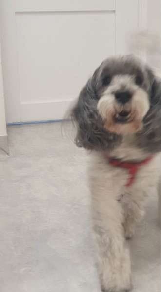
A while ago, an 11-year-old female dog named Emma came to our clinic; for about two weeks she had been urinating more frequently on walks. Her owner was worried by this behaviour, because Emma had previously had bladder infections, so she came to us straight away with the results of a freshly performed urine test – which was itself a very good decision, since it sped up the whole diagnostic process. The test indicated inflammation, and the urine sediment also contained calcium oxalate crystals as well as struvite. For us, as a team, that result immediately suggested we were dealing with something more than a passing bladder irritation – the presence of two different types of crystals at once was a signal that something in Emma's urinary tract needed a closer look.
A Surprisingly Calm Patient
Emma was in good spirits at the time. She had an appetite, was happy to go on walks, and showed no signs of pain or discomfort. During the clinical examination, almost everything appeared normal – she behaved as if nothing serious was really going on. That in itself is valuable clinical information: an animal that's weak, feverish, or losing its appetite immediately raises more concern and speeds up diagnostic decisions, whereas a patient in good general condition can easily lull both the owner and, without further tests, the vet into a false sense of security.
Hard as Stone – Clinical Examination of the Bladder
Everything changed the moment Wiktoria Król, DVM, began examining the bladder through the abdominal wall. Something was clearly wrong, because it was very hard – quite unlike the normal, urine-filled bladder examined on a daily basis. Even so, Emma kept standing bravely on the table and didn't even flinch, even though this kind of pressure should have been clearly painful had it been a classic bladder infection. This contrast – an organ hard as stone with a complete absence of a pain response – was the first strong signal that the situation was more complex than the history alone suggested.
An Ultrasound That Explained Nothing
The next step was an abdominal ultrasound (USG) focused on the bladder. Here we ran into an obstacle: the entire bladder was impossible to assess properly, because something was blocking the view and preventing a clear image of its interior and wall. Instead of clarifying the clinical picture, the ultrasound complicated it further – we still didn't know what was causing Emma's frequent urination, though we now knew for certain that something unusual was inside the bladder.
X-ray and a Stone Measuring 64 by 76 Millimetres
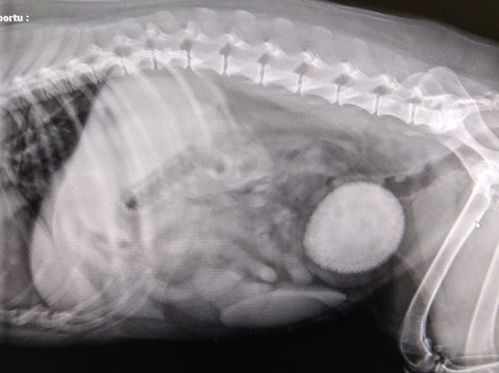
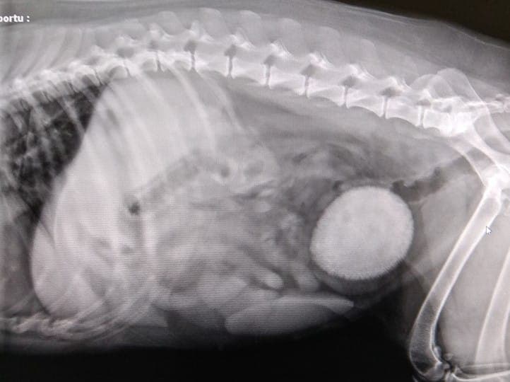
It was only the X-ray that made the whole picture clear. The image showed the entire bladder taken up by a single stone measuring roughly 64 by 76 millimetres – a structure that, in practice, filled almost the whole organ. It was hard to believe that such an enormous stone could have been forming for a long time without producing any classic pain-related symptoms, only a discreet, easy-to-dismiss increase in urination frequency.
Emma's story is a good reminder of a simple but often underestimated rule: an animal's lack of pain and good overall spirits don't mean there's no serious problem in the urinary tract. Bladder stones can grow slowly and silently, and the only signal may be a subtle change in urination habits – which is why any such change is worth reporting to a vet, even if the dog looks and behaves completely normal.
What Is Urolithiasis (Urinary Stones) in Dogs
How Urinary Stones Form
Urinary stones form as a result of the crystallisation of mineral salts present in urine. Most often we're talking about calcium oxalate, calcium phosphate, urates, cystine compounds, and above all ammonium magnesium phosphate, or struvite, which is one of the most frequently recognised types of stone in dogs. Under the right conditions, these compounds begin to precipitate out of solution, and once there's enough of them, they first form tiny crystals, then larger clusters and deposits, and finally fully formed stones. The exact mechanism behind stone formation still isn't fully understood – it depends on multiple factors acting at once, such as the rate at which urine flows and sits in the bladder, the concentration of individual salts, the presence of a so-called crystallisation nucleus (a microscopic point around which further layers of mineral start to accumulate), and sometimes the presence of bacteria that favour the formation of certain types of stone.
The Role of Urine pH
It's worth remembering that the urine of carnivores is normally acidic. Any shift in that pH can favour the crystallisation of particular compounds. In urine that's neutral to alkaline, struvite tends to precipitate more readily, while in urine that's acidic to neutral, calcium oxalate, urates, cystine compounds, or silicates tend to form instead. This is why diet choices for preventing and treating urinary stones are so closely tied to regulating urine pH – shifting the pH in one direction can help dissolve one type of stone while, at the same time, favouring the formation of a completely different one if the diet isn't chosen correctly and isn't supervised by a vet.
Bladder Stones versus Stones in the Kidneys, Ureters and Urethra
Mineral deposits can form in different parts of the urinary tract, and their location matters a great deal for the clinical picture. Stones sitting in the bladder, as in Emma's case, usually cause symptoms related to urination itself – frequent urination, discomfort, sometimes blood in the urine – although, as this case shows, they can also remain silent for a long time. Stones in the kidneys can stay clinically silent for many months and are sometimes found incidentally during imaging performed for a completely different reason. The most dangerous situation arises when a stone moves and blocks a ureter – the tube connecting the kidney to the bladder – which can lead to serious damage to the kidney on the blocked side. Stones that reach the urethra, meanwhile, can cause partial or complete obstruction of urine outflow, which in dogs, especially males, can be an emergency requiring immediate intervention.
Types of Urinary Stones in Dogs
Struvite
Struvite – ammonium magnesium phosphate deposits – is one of the most frequently recognised types of urinary stone in dogs. It usually forms in urine that's neutral to alkaline, and its formation is very often encouraged by the presence of urease-producing bacteria, an enzyme that breaks down urea and raises urine pH. That's one reason why struvite stones in dogs (unlike in cats, where they more often form in sterile urine) are fairly closely linked to bacterial urinary tract infections. The good news is that struvite can often be dissolved using an appropriately chosen therapeutic diet, combined – if an infection has been confirmed – with antibiotics, under regular monitoring by a vet with follow-up imaging.
Calcium Oxalate
Calcium oxalate is the second very commonly recognised type of stone in dogs, and it was precisely this type, alongside struvite, that was found in Emma's urine. Unlike struvite, this kind of stone generally does not dissolve with diet – once a calcium oxalate stone has formed, it remains a stone, and the only way to remove it is surgery or, at some referral centres, other minimally invasive techniques. An appropriately chosen diet mainly plays a preventive role here – it reduces the risk of further stones forming in the future, but it won't remove an existing one.
Urate Stones
Urate stones occur less often than struvite and calcium oxalate, but they're well known in veterinary medicine, especially in breeds predisposed due to their particular purine metabolism – a classic example being Dalmatians and English Bulldogs. In these breeds, an altered way of processing uric acid increases the risk of urate stones forming even on an apparently normal diet, which is why dogs of these breeds, especially those with a family history of urinary problems, are worth watching particularly closely.
Cystine Stones
Cystine stones are linked to a defect in the kidneys' reabsorption of the amino acid cystine – a condition known as cystinuria, which has a genetic basis and occurs considerably more often in males than in females. Among the breeds described as predisposed to this type of stone are Dachshunds, among others. Like calcium oxalate, this type of stone usually doesn't dissolve with diet alone and most often requires surgical removal, although long-term dietary management and adequate hydration can significantly reduce the risk of recurrence.
Why the Composition of the Stone Matters
Emma's story illustrates well why knowing the exact composition of a stone matters so much. Until you know what a stone is made of, it's hard to plan conservative treatment or prevention with any real confidence – a diet designed to dissolve struvite won't help a stone made mainly of calcium oxalate, and vice versa. That's why every surgically removed stone should, whenever possible, be sent for laboratory analysis. Only the result of that analysis makes it possible to plan a diet and a monitoring schedule that genuinely reduce the risk of the problem coming back.
Causes and Risk Factors for Urinary Stones
Diet and Feeding Habits
The way a dog is fed has a direct effect on the mineral composition and pH of its urine, and therefore, indirectly, on the risk of different types of stones forming. That's one reason why choosing food for a dog with a history of urinary problems should be discussed with a vet, rather than decided independently based on opinions found online or other owners' experiences.
Insufficient Water Intake
The more concentrated the urine, the easier it is for it to become supersaturated with minerals and start the crystallisation process. Dogs that drink too little water – for various reasons, from a diet based solely on dry food to simply having a lower natural inclination to drink – produce more concentrated urine, which favours stone formation. Increasing water intake is one of the simplest, and at the same time one of the most effective, elements of prevention.
Excess Weight and Low Physical Activity
Overweight, less active dogs urinate less often and less regularly, which lengthens how long urine sits in the bladder. Longer contact between urine and the bladder wall, combined with other risk factors, can favour mineral crystallisation and the development of secondary infections. Maintaining a healthy body weight and regular exercise are therefore not just a matter of general fitness, but a real element of urinary prevention.
Breed Predispositions
Certain breeds and bloodlines show a clearly greater tendency towards specific types of urinary stone – Dachshunds towards cystine stones, Miniature Schnauzers towards struvite and calcium oxalate, Dalmatians and English Bulldogs towards urate stones. It's worth remembering, though, that urinary stones can affect a dog of any breed and any size, including mixed breeds – much like the case of Franek the cat from our practice, described elsewhere, where the problem affected an ordinary mixed-breed cat. Among dogs, too, it's not worth assuming "this doesn't apply to us" just because a dog doesn't belong to a breed considered more predisposed.
Urinary Tract Infections
Bacterial infections, especially those caused by urease-producing bacteria, can directly encourage the formation of struvite stones, while at the same time producing symptoms very similar to those of urinary stones themselves – frequent urination, discomfort, sometimes blood in the urine. That's why a urine test, together with a bacterial culture if needed, is an important part of full diagnostics, regardless of whether a stone is already visible on imaging or not.
Sex and the Anatomy of the Urethra
The anatomy of the urinary tract differs significantly between females and males, which has direct clinical significance. Females have a relatively shorter, wider urethra, so even a sizeable stone in the bladder, as in Emma's case, may fail to cause obstruction for a long time, although of course it can still produce other symptoms or, as here, hardly any symptoms beyond a discreet increase in urination frequency. In males, the urethra is narrower and longer, which means that even small stones can more easily lodge inside it, leading to partial or complete obstruction of urine outflow – a situation far more urgent and dangerous than the mere presence of a stone in the bladder.
Age and General Health
Urinary stones are sometimes associated exclusively with younger animals, but Emma's case, an 11-year-old female dog, shows that the problem can build up over years and only reveal itself later in life. In older dogs there's an added risk linked to coexisting conditions – hormonal disorders, chronic kidney disease, or a general slowdown in metabolism – which can affect the composition and concentration of urine. That's why, in older dogs with urinary symptoms, vets often broaden the diagnostic work-up to include an assessment of overall health rather than focusing solely on the bladder itself. It's worth stressing that age alone isn't a contraindication for stone-removal surgery – what matters most is the patient's overall health, not the number of years. Emma was 11 years old and, despite complications related to the size of the stone, tolerated the surgery very well and made a quick recovery. In older dogs, vets typically broaden pre-operative testing to include an assessment of heart, kidney, and liver function, in order to estimate anaesthetic risk as accurately as possible and plan the procedure and post-operative care accordingly.
Symptoms That Should Concern an Owner
Symptoms of urinary stones can vary, and they don't always all appear together. The following signs should prompt a visit to the vet:
- frequent urination – more frequent trips outside than usual, often passing only a small amount of urine each time,
- painful urination, visible straining, or urgency,
- passing urine in small "dribbles" instead of a normal, continuous stream,
- blood in the urine – urine tinged pink, red, or clearly bloody,
- urinating in unusual places, especially if the dog was previously fully house-trained,
- excessive licking around the urethral opening,
- lethargy, reduced appetite, and in some cases vomiting,
- in extreme cases – a complete inability to urinate despite repeated straining.
It's worth remembering, though – as Emma's story shows – that even a fairly large stone in the bladder can go without any classic clinical symptoms for a long time, aside from a subtle change in urination frequency. That's why paying close attention to a dog's everyday habits matters just as much as reacting to obvious pain or blood in the urine.
Complete obstruction of the urinary tract is always an emergency, life-threatening condition, whether in a dog or a cat. If a dog repeatedly and unsuccessfully attempts to urinate, is clearly restless, and no urine is being produced, the situation calls for immediate veterinary help – ideally that same day, not "wait and see until tomorrow". Untreated obstruction can quickly lead to serious systemic complications.
How Urinary Stones Are Diagnosed in Dogs
History and Clinical Examination
Diagnosis always starts with a thorough history – when the symptoms began, how often the dog urinates, whether the diet has changed, whether the dog is drinking more or less than usual, and whether similar problems have occurred before. The vet then carries out a clinical examination, including careful palpation of the abdomen, which – as in Emma's case – can sometimes reveal an abnormally hard or enlarged bladder.
Urinalysis
A general urine test is usually the first diagnostic step, since it provides a great deal of valuable information: the urine's pH, its specific gravity, and above all what's present in the sediment. Urine sediment can contain various types of crystals, epithelial cells, inflammatory cells, bacteria, and erythrocytes (red blood cells). In Emma's case, it was precisely this test, performed before the visit, that was the first to signal the presence of inflammation and two different types of crystals.
Bladder Ultrasound
When the urine test result is concerning, the next step is usually an ultrasound (USG). It allows the vet to assess the appearance of the bladder wall – whether it's thickened, whether the urine is clear, or whether "floating" crystals or formed stones are already visible. As in Emma's case, though, ultrasound can be difficult to interpret conclusively if something is obscuring the view of the bladder or if there's very little urine in the organ – in such situations, repeating the scan or turning to another imaging method may be necessary.
X-ray of the Urinary Tract
An abdominal X-ray can be an invaluable complement to ultrasound, especially when the ultrasound picture is ambiguous, as it was for Emma. Many types of stone, including struvite and calcium oxalate, show up well on a standard X-ray, which makes it possible to confirm their number, size, and location. It's worth knowing, though, that some types of stone – for example, some urate and cystine stones – can be less visible on a plain X-ray, which sometimes makes contrast studies or other imaging techniques necessary.
Blood Tests and Urine Culture
Depending on the clinical picture, the vet may also recommend blood tests to assess overall health and kidney function, along with a urine culture with antibiotic sensitivity testing, especially if a concurrent bacterial infection is suspected. These tests aren't always necessary in every case, but they're a valuable tool when the clinical picture is more complex or treatment isn't producing the expected improvement.
Treatment of Urinary Stones
Conservative Treatment – Dissolving Stones Through Diet
For some types of stone, above all struvite, conservative treatment with a specialised therapeutic diet, available only on veterinary recommendation, is sometimes possible, often combined with antibiotics if a bacterial infection has been confirmed. This approach requires patience – it usually takes several weeks to a few months – along with regular check-ups, most often with repeat imaging, to monitor whether the stone is actually shrinking.
Surgical Treatment – Cystotomy
Cystotomy, the surgical opening of the bladder to remove stones, is a procedure routinely performed in small-animal surgery, although – as we noted when describing Emma's case – it doesn't happen every day. In her case, treatment involved exactly that: surgically removing the stone from the bladder, because with such a large stone, whose exact composition wasn't yet known at the time, there was no other reasonable course of action. Despite the complications involved, Emma coped with the surgery very bravely.
Recovery After Surgery
After a cystotomy, a dog usually wears a protective collar for a while to prevent licking at the stitches, and the vet sets a schedule for checking the wound's healing and confirming that the animal is urinating normally. In the first few days after surgery, it's worth limiting intense physical activity – long walks, jumping, and play with other dogs should wait until the wound has healed. Emma recovered surprisingly quickly, which shows that even major surgery in an older dog can have a very good outcome if it's carried out at the right time.
Laboratory Analysis of the Removed Stone
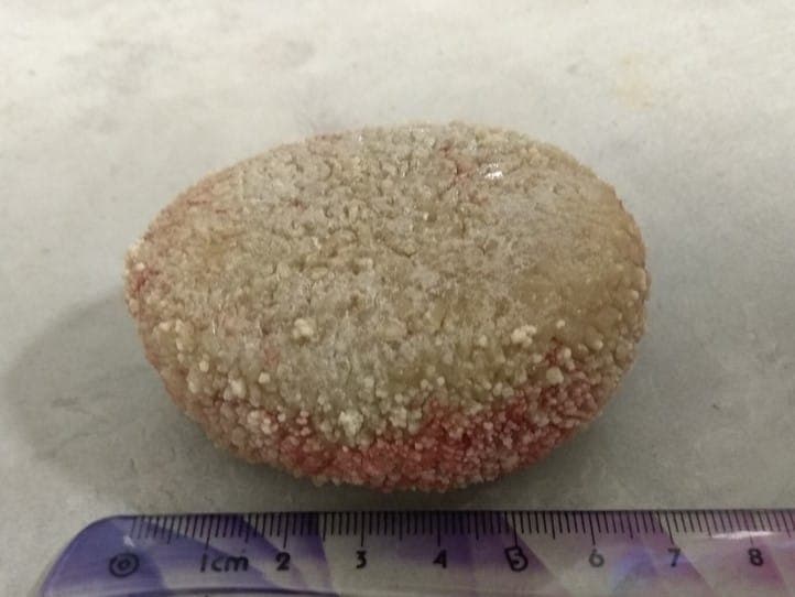
We sent the stone removed from Emma's bladder for chemical analysis to the Minnesota Urolith Center in the United States – one of the reference facilities that studies this type of deposit. Unfortunately, that means it didn't stay with us at the clinic as a keepsake. All we have left is the photo, which – as we like to point out – should serve more as a cautionary tale for other owners than as something to boast about.
Preventing Recurrence
Preventive Diet and Regular Check-ups
In mild cases of crystalluria – meaning the presence of crystals in the urine without any stones having formed yet – a change of diet and appropriate supplements to support a healthy urine pH are often enough. What matters a great deal, though, is that in such situations the urine needs to be checked regularly, brought in for testing according to the vet's recommendations, even if the dog seems to be feeling fine.
Encouraging Your Dog to Drink Water
Since concentrated urine favours mineral crystallisation, increasing the amount of water a dog drinks is one of the simplest and most effective elements of prevention. This can be helped by:
- incorporating more wet food into the diet, unless the vet advises otherwise,
- placing several bowls of fresh water in different spots around the house,
- changing the water regularly and keeping bowls and containers clean,
- encouraging drinking during and after walks, especially on warm days.
Maintaining a Healthy Body Weight
Maintaining a healthy weight and regular physical activity – walks, play, running – support the overall health of the urinary tract, improve the regularity of urination, and reduce the risk of urine sitting in the bladder for too long.
Why Urinary Stones Tend to Recur
Urinary stones are a condition with a clear tendency to recur. Many owners assume that once the problem has been treated – whether through diet or surgery – there's no need to keep worrying about it. That mistaken belief is one of the most common causes of further, sometimes more serious, episodes. Regular follow-up urine tests, and sometimes ultrasounds too, make it possible to catch a recurrence at an early, easier-to-treat stage, before full clinical symptoms develop.
Frequently Asked Questions
Do urinary stones always hurt?
Not always. Emma's story shows that even a large stone filling almost the entire bladder can go without obvious pain-related symptoms for a long time. That's why it's worth paying attention to subtle behavioural changes, such as slightly more frequent urination, rather than only to obvious pain or blood in the urine.
Does every urinary stone require surgery?
Not always. Struvite stones, especially when accompanied by a bacterial infection, can often be dissolved with an appropriately chosen therapeutic diet under veterinary supervision. Calcium oxalate, cystine, and urate stones usually don't dissolve with diet and require surgical removal or other minimally invasive techniques. The decision is always made by the vet based on the full clinical picture and, whenever possible, knowledge of the stone's composition.
How long after treatment should urine be monitored?
The frequency of check-ups is set individually by the treating vet, depending on the type of stone, the treatment used, and the dog's medical history. The general rule, though, is that monitoring shouldn't stop just because symptoms have resolved – urinary stones have a tendency to recur, and regular testing helps catch the problem before it makes itself clearly known again.
Which dog breeds are more prone to urinary stones?
Certain breeds really do show a greater tendency towards particular types of stone – Dachshunds towards cystine stones, Miniature Schnauzers towards struvite and calcium oxalate, Dalmatians and English Bulldogs towards urate stones. It's worth remembering, though, that urinary stones can occur in a dog of any breed and size, including mixed breeds, so watching your pet's urination habits makes sense regardless of its breed.
What should I do if my dog can't urinate at all?
This calls for immediate veterinary help, ideally that same day. Complete obstruction of the urinary tract is a life-threatening condition, especially in males, whose narrower urethra is more easily blocked by stones.
Is a preventive diet enough to avoid recurrence?
An appropriate diet is a very important, but usually not the only, part of prevention. Increasing water intake, maintaining a healthy body weight, and regular follow-up urine tests also matter a great deal, especially in the period right after treatment ends.
Are urinary stones hereditary?
For some types of stone, for example cystine stones linked to cystinuria, genetic predisposition plays a significant role, which is why certain bloodlines and breeds are more burdened by them than others. In most cases, though, urinary stones result from the interplay of many factors at once – diet, hydration, infection, individual anatomy – rather than heredity alone.
Emma's story shows just how misleading it can be to rely solely on the presence or absence of pain when assessing a dog's urinary health, and how much patient, multi-step diagnostics – urinalysis, ultrasound, X-ray – contribute before deciding on further treatment. If you notice your dog urinating more often, blood in the urine, straining while urinating, or any other worrying sign involving the urinary tract, don't wait for it to sort itself out. At the Hau-Miau clinic at ul. Siemieńskiego 23 in Warsaw's Ochota district, we've spent years diagnosing and treating urinary tract conditions in dogs and cats, combining clinical examination, ultrasound, X-ray, and laboratory testing into one coherent diagnostic path. Book a visit — call +48 22 823 35 63.