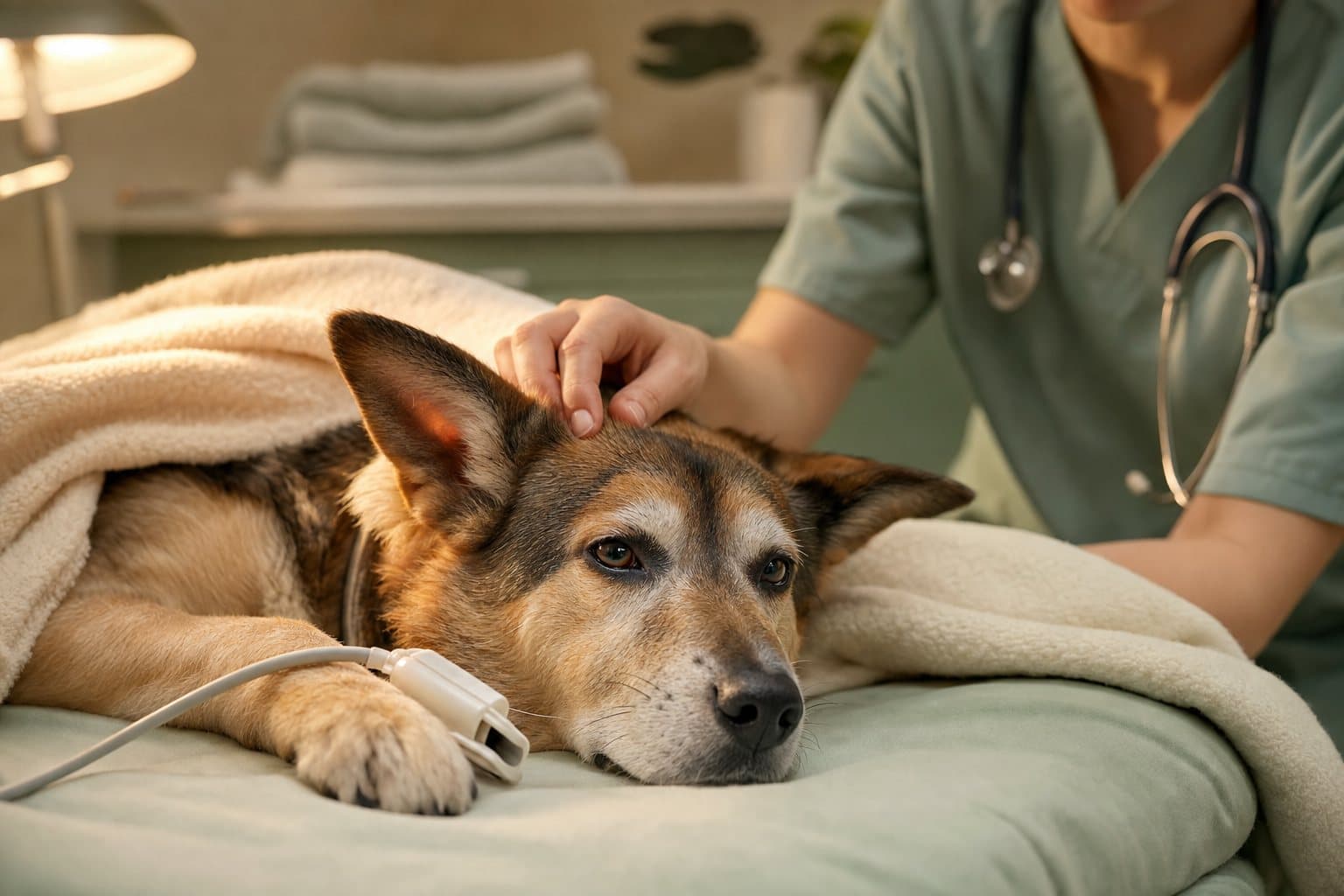
Hearing the word "surgery" from your vet is unsettling, even when you know it's the only path back to health for your pet. Sometimes an illness does not respond to conservative treatment, and surgical intervention is required. Spleen tumours, for example, call for exactly this approach – the entire organ is removed, and the animal typically makes a full recovery, because a dog's or cat's body copes well without it. At the Hau-Miau clinic at ul. Siemieńskiego 23 in Warsaw's Ochota district, we've spent years performing soft tissue surgery – from simple, planned operations to life-saving emergency interventions. In this article we explain when surgery becomes necessary, which types of operations we perform most often, what our operating theatre looks like, and what safety standards we apply at every stage of treatment – from the first diagnosis, through anaesthesia, to the first hours after your pet wakes up. We've tried to describe all of this as concretely as possible, without unnecessary alarm, because a clear picture of what's ahead usually reduces the stress of the visit itself – for the animal and the owner alike.
When Surgery Is Necessary
When Conservative Treatment Isn't Enough
Most of the conditions you bring to our clinic can be managed with conservative treatment: medication, diet, a change in lifestyle, sometimes physiotherapy. We turn to surgery only when a problem is mechanical or structural in nature – the kind no tablet can fix. A tumour blocking the intestine, a stone obstructing the urethra, a twisted stomach, a broken bone, a hernia, an infected uterus – in these situations, delaying the decision to operate usually worsens the outlook rather than buying the animal more time. Whether a given animal needs surgery is always a decision the vet makes individually, based on a clinical examination and diagnostic tests. Every patient, every tumour, and every hernia looks different, so there's no room here for comparisons with a "similar case" found online, or for deciding on your own whether surgery can still wait.
Spleen Tumours and Splenectomy: An Example From Our Practice
A good example is splenectomy, the complete surgical removal of the spleen. Spleen tumours in dogs, and less often in cats, are dangerous less because of their mere presence and more because of the risk of rupture and bleeding into the abdominal cavity – an emergency, life-threatening state requiring immediate intervention. The spleen isn't an organ essential for survival: once it's removed, other organs, chiefly the liver and bone marrow, take over part of its role in filtering blood and supporting immunity, and the animal typically returns to normal function. This procedure, though serious and requiring an abdominal incision, is performed routinely in small-animal surgery.
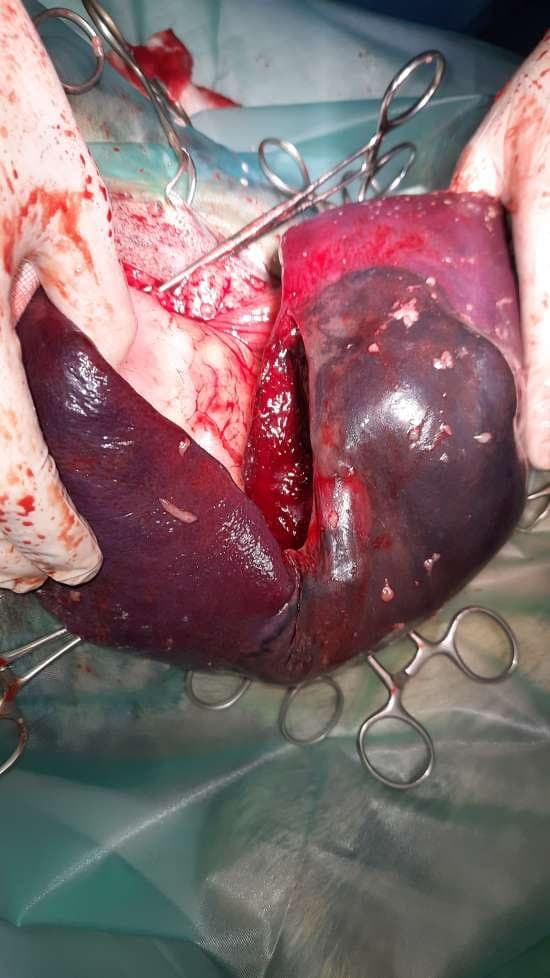
The removed organ, or a sample taken from it, is usually sent for histopathological examination. Only that result allows us to say with confidence what kind of growth we were dealing with – benign or malignant – and to plan any further treatment that might be needed. Without this test, the surgeon and owner would be working in the dark, relying solely on how the tumour looks to the naked eye, which is never enough in veterinary oncology.
The Role of Diagnostics Before Deciding on Surgery
Before a decision to operate is even made, an animal usually goes through a set of tests that help establish whether surgery is necessary and whether the patient can safely withstand it. Depending on the case, this might include an abdominal ultrasound, an X-ray, blood tests (a complete blood count and biochemistry panel), and a urinalysis. Ultrasound lets us see internal organs, their structure, and any focal changes; X-rays show up bones, some urinary stones, and changes in the chest particularly well; and blood and urine tests reveal how the kidneys, liver, and other organs are functioning before we even start planning anaesthesia. It's this diagnostic picture, not a mere suspicion of illness, that ultimately determines whether surgery is indicated, how urgent it is, and how extensive it needs to be.
Sometimes an imaging result is ambiguous and needs to be repeated or supplemented with another method – that's a normal part of thorough diagnostics, not a sign that something has gone wrong. It's better to repeat a test and be certain than to decide to open the abdominal cavity on the basis of an incomplete picture.
Planned Procedures vs. Emergency Procedures
In our practice we distinguish between two broad categories of surgery. Planned procedures are the ones there's time for: castration, removal of a skin growth, oral cavity treatment with tooth extraction. These are preceded by full diagnostics, a conversation with the owner, and the setting of a convenient date, so that both the animal and the household are prepared for the day. Emergency procedures are an entirely different situation – a road accident, a fracture, a complicated bite wound, an acute abdomen, a twisted stomach. Here, time is everything, and decisions are made in minutes, not days or hours.
Both types of surgery require the same infrastructure – an operating theatre, anaesthesia, and a qualified team – but a completely different pace of decision-making. A planned procedure gives us the comfort of fully preparing the patient beforehand; an emergency procedure often means operating on an animal that arrives on the table already weakened, so stabilisation and the surgery itself have to proceed side by side.
The Pre-Surgical Consultation for Planned Procedures
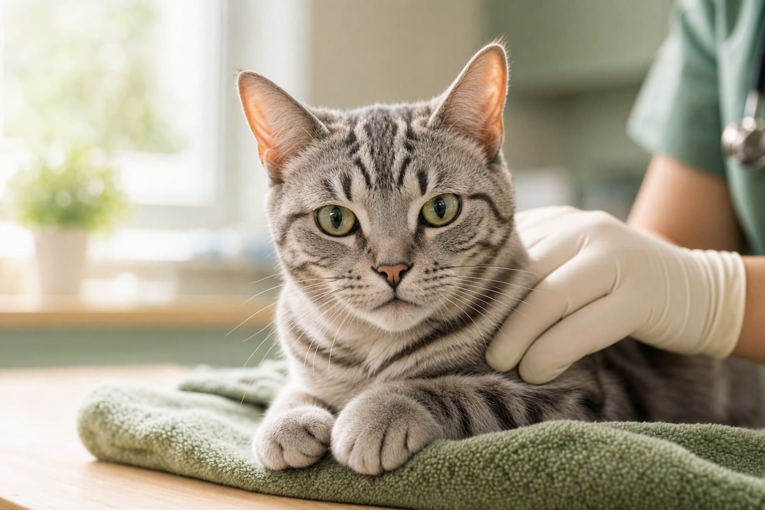
Before we schedule a planned operation, we invite the owner and the pet in for a qualifying consultation. The vet carries out a full clinical examination, asks about past illnesses, current medication, and any previous reactions to anaesthesia (if the animal has been operated on before), as well as any worrying symptoms noticed recently. This is also the moment to ask every question on your mind: how long your pet will stay at the clinic, what kind of recovery to expect, whether inhalation anaesthesia is recommended in this particular case. Based on that conversation and the results of any additional tests, the vet decides whether the animal can be scheduled for the nearest available date, or whether some health parameter – heart or kidney function, say – needs to be stabilised first.
Types of Surgical Procedures We Perform
Below we describe the most common types of procedures we carry out in our operating theatre. The vast majority fall under what's known as soft tissue surgery.
What Is Soft Tissue Surgery
Soft tissue surgery covers procedures on everything except bones and joints: the skin, muscles, organs of the abdominal and thoracic cavities, and the urogenital system. It's a distinct field from orthopaedic surgery, which deals with fractures, dislocations, and ligament reconstruction, and usually calls for different equipment and a different specialisation. Our operating theatre is set up chiefly for soft tissue procedures – from simple interventions lasting a matter of minutes to complex, multi-hour abdominal operations.
What all of these procedures share is that they involve well-perfused, injury-prone soft organs, which presents the surgeon with different challenges from working on bone. Handling tissue with precision, limiting the time an organ stays exposed, and carefully controlling bleeding matter just as much here as cutting and suturing technique itself. That's why a surgeon's experience – measured in years of practice and hundreds of procedures performed – has a real bearing on how long an operation takes and how quickly the patient recovers.
Surgery on the Digestive System
Animals come to our clinic after swallowing a foreign object – a piece of a toy, a bone, a scrap of fabric or string – that has become lodged in the stomach or intestine. In such cases we perform a gastrotomy or enterotomy: a surgical opening of the stomach or intestine, removal of the obstruction, and careful closure of the organ with sutures. The same type of procedure is used for narrowing of the intestinal lumen, for perforation (a hole in the wall), and for a tumour blocking the passage of food.
A separate, serious group of interventions is gastropexy, a procedure in which the stomach is surgically anchored to the inner abdominal wall. It's performed most often in large dogs of breeds predisposed to gastric torsion, known by the abbreviation GDV – a condition in which the stomach rotates around its own axis, cutting off its own blood supply and trapping gas inside. This is one of the few conditions in veterinary medicine where every hour truly counts, and without immediate surgery the animal has very little chance of survival.
Gastric torsion (GDV) is an emergency. A tense, distended, drum-like abdomen, unproductive retching, restlessness, drooling, and weakness in a large-breed dog are signs to get to the clinic immediately – don't wait until morning, and don't hope it will pass on its own.
Surgery on the Urogenital System
The second large group of procedures we perform involves the urogenital system. These include repairs of inguinal and abdominal hernias, removal of the uterus in diseases such as pyometra or tumours, and the removal of stones and abscesses from the bladder. In an inguinal hernia, the surgeon opens the inguinal canal, returns the displaced organs to the abdominal cavity, and closes the defect that allowed the displacement in the first place. An abdominal hernia, in turn, requires opening the abdominal wall to return the organs to their proper position.
The most frequently performed procedure in this group, though, remains castration, which we cover in more detail in the next section, since it's the source of the most questions – and terminology mix-ups – in conversations with owners.
Castration vs. Sterilisation: An Important Distinction
At our clinic we perform castration, not sterilisation, although in everyday conversation the two words are often used interchangeably. Castration is a procedure in which the testicles are removed in males, and the ovaries together with the uterus in females – after the procedure, the animal no longer has hormonally active gonads. Sterilisation is a different procedure: the gonads (testicles or ovaries) stay in place, and only the reproductive pathways are severed – the vas deferens in males or the fallopian tubes in females. The practical effect is similar (the animal can no longer reproduce), but the scope of the procedure itself and its hormonal consequences for the body are different.
This distinction has practical implications, because some of the diseases castration helps prevent – certain mammary tumours, pyometra, and prostate disease among them – are linked to the long-term action of sex hormones, not merely to the capacity to reproduce. If you're unsure which option, and at what age, is right for your particular pet, we encourage you to discuss it with the treating vet at the qualifying visit.
Oncological Surgery: Tumours and Skin Growths
When a tumour appears in the skin or subcutaneous tissue, the decision to remove it always follows a clinical assessment, and often a prior biopsy, which helps estimate what we're dealing with before the procedure. If a growth raises suspicion of malignancy, the surgeon removes it with a margin of healthy surrounding tissue, known as an oncological margin, to reduce the risk of leaving cancerous cells behind in the body. The removed material is then, almost without exception, sent for histopathological examination – the only reliable way to determine what type of growth it was, assess whether the excision margin was sufficient, and plan whether further treatment or monitoring is needed.
Not every skin growth that brings you to our clinic is a tumour. Among the most commonly removed lesions are benign lipomas, adenomas, and warts, which a surgeon typically removes when they bother the animal, are prone to mechanical injury (for example, from a collar or from scratching), or change in appearance quickly. Whether a particular growth needs prompt removal, or can simply be monitored, is always a decision made by the vet on examination, often supported by a fine-needle biopsy.
Abscesses, Cysts, and Chronic Non-Healing Wounds
A separate group of soft tissue procedures involves abscesses, cysts, and wounds that refuse to heal despite the passage of time. An abscess is a closed pocket of pus, most often the result of an infection, for instance following a bite from another animal – surgically opening, cleaning, and draining it usually brings much faster improvement than antibiotics alone. Cysts, closed structures filled with fluid or tissue material, are removed surgically when they grow, rupture repeatedly, or interfere with the animal's normal functioning. Chronic wounds that fail to heal despite dressings and topical treatment often need surgical debridement – removing dead tissue and re-closing the wound in theatre conditions, rather than continuing to wait for it to heal on its own.
Tooth Extractions and Oral Cavity Treatment
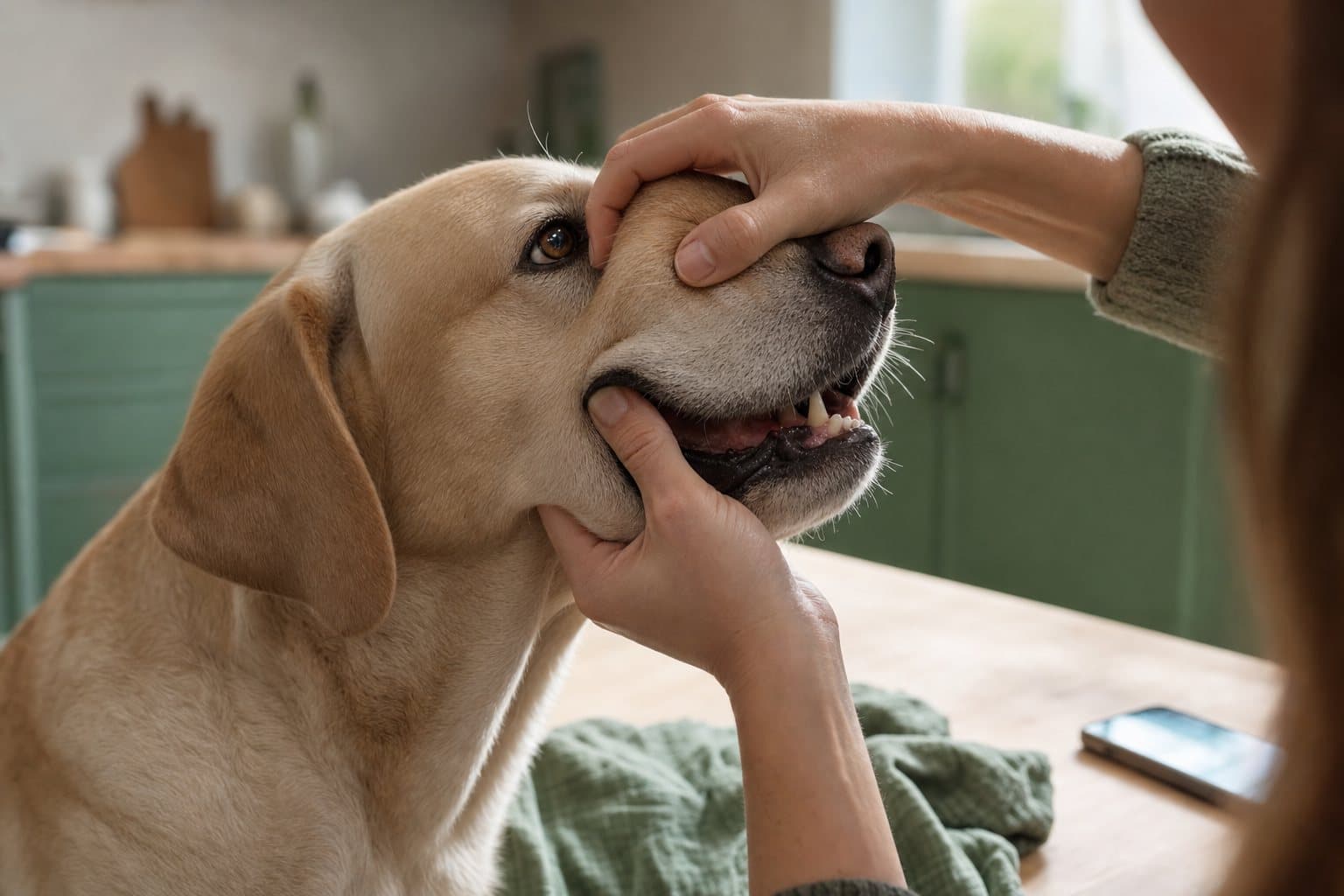
Surgery isn't limited to the abdomen. Tooth extraction – the surgical removal of a tooth – is one of the more commonly performed procedures in older dogs and cats with advanced periodontal disease. Teeth affected by chronic inflammation are often a source of pain the animal lives with daily without showing it clearly, as well as a focus of infection that places a burden on the whole body, with knock-on effects, as clinical observation suggests, for heart and kidney health over the longer term. Removing them, though it sounds drastic, genuinely improves the patient's quality of life – owners very often notice their pet returning to its old activity levels and appetite within just a few days of the procedure, sometimes tackling dry food more readily than it did right before treatment, when eating hurt.
Emergency Procedures
Emergency procedures include operations following road accidents, fractures requiring surgical fixation, complicated bite or tear wounds, and some respiratory and circulatory conditions where restoring normal organ function requires a surgeon's intervention. In contaminated wounds, the surgeon must first thoroughly clean the tissue of dead material and debris before placing sutures. Skipping this step, even in the name of closing the wound faster, usually ends with the wound failing to heal properly and the risk of infection rising sharply. In road accident cases, surgery is often preceded by intensive stabilisation of the patient – correcting circulation, oxygen therapy, pain relief – because an animal in shock isn't a good candidate for immediate general anaesthesia, unless the condition is so severe that the risk of further delay outweighs the risk of the procedure itself.
The Surgical Theatre and Safety Standards
Operations are carried out in a dedicated surgical theatre, which is disinfected with UV light every night. This isn't a general-purpose room used for one procedure one day and something else entirely the next – its sole purpose is surgery, which lets us maintain a high standard of cleanliness and reduce the risk of hospital-acquired infection.
Isoflurane Inhalation Anaesthesia
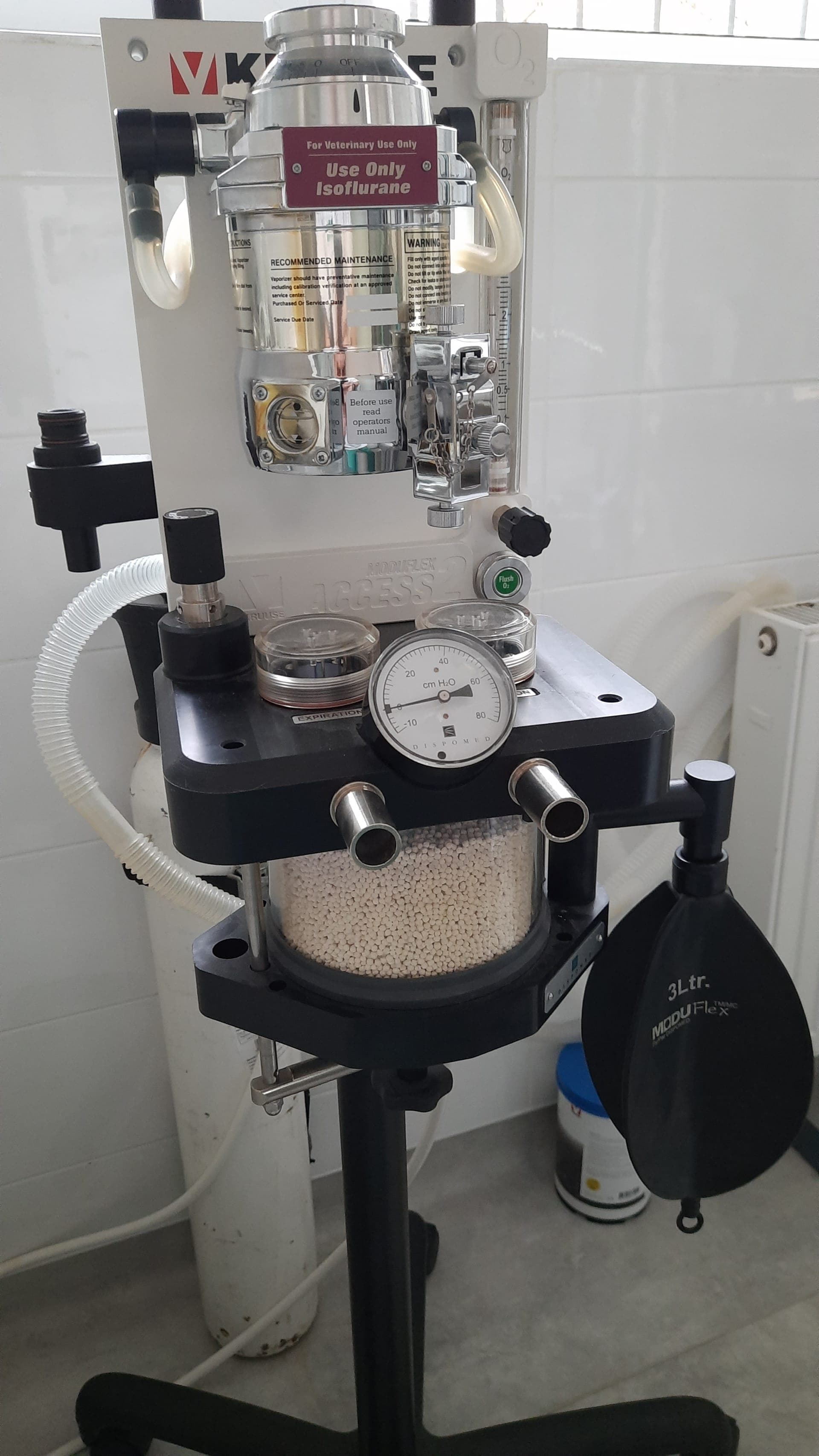
Our theatre is equipped with an isoflurane inhalation anaesthesia machine. Inhalation anaesthesia differs from traditional (infusion) anaesthesia in that, instead of medication administered intravenously, a mixture of gas (isoflurane) and medical oxygen – which the animal breathes throughout the operation – is used to maintain sleep during the surgical procedure. The anaesthetist adjusts the gas dose continuously, matching the depth of anaesthesia to how the procedure is progressing and how the patient is responding.
Why We Recommend It for Higher-Risk Patients
We recommend this type of general anaesthesia especially for brachycephalic (short-skulled) breeds, such as pugs and French or English bulldogs, for animals with additional health conditions, and for older patients, due to the lower burden anaesthetics place on the body. Isoflurane's hepatic metabolism is only around 0.25%, meaning the gas is exhaled in practically unchanged form and places very little strain on the liver, unlike some intravenous medications, which the body has to break down metabolically. For a patient whose liver already has its hands full – because it's ill, elderly, or has an unusual airway structure that makes breathing harder – that difference has a real bearing on the safety of the entire procedure.
Local Anaesthesia and Sedation
Not every procedure calls for full general anaesthesia. Some minor procedures – treating a superficial wound, removing a single small skin lump, or certain diagnostic tests – can be carried out under sedation, sometimes combined with local anaesthesia of the surgical site. Sedation lowers the animal's level of awareness and stress and provides pain relief, but it doesn't suppress reflexes as deeply as full anaesthesia does. Whether a given procedure can be done under sedation, or requires full general anaesthesia with intubation and ventilatory support, is assessed case by case by the vet, taking into account the scope of the procedure, the animal's temperament, and its state of health.
Anaesthesia Care During the Procedure
Dedicated anaesthesia care ensures patient safety throughout the procedure. The person responsible for monitoring anaesthesia observes the animal continuously, from premedication through induction and maintenance of anaesthesia to full recovery. This is a distinct role from the surgeon's, who can focus entirely on the surgical field during this time, without simultaneously worrying about the patient's vital signs.
UV Disinfection of the Theatre
UV light is used in medicine to reduce the number of microorganisms on surfaces that are hard to reach with cleaning agents alone. The surgical theatre is disinfected every night, after all of that day's procedures are finished, so that by the next morning the room is ready to receive the next patient in conditions as close to sterile as possible.
Suture Materials and Wound Closure
The surgeon chooses how to close a wound based on the type of tissue and the location of the procedure. Internal organs and deeper tissue layers are typically closed with absorbable sutures, which the body gradually breaks down on its own, so they never need to be removed later. Skin may be closed with non-absorbable sutures, surgical staples, or an intradermal (buried) suture, depending on the procedure and the surgeon's preference. If non-absorbable sutures were used, a follow-up visit will be needed to remove them, usually a couple of weeks after surgery, once the wound has healed well.
Minimising Perioperative Risk
Surgical safety is the sum of many small elements, not one single "main" factor. It's made up of thorough pre-operative diagnostics, an anaesthesia method matched to the individual patient, the constant presence of someone monitoring vital signs during the procedure, work carried out in a dedicated, regularly disinfected theatre, and post-operative observation before the animal goes home. No single element can substitute for the others – it's their combination that makes a procedure that sounds frightening a routine part of clinical practice. The owner's side of preparation matters too – proper fasting before the procedure and following post-operative instructions reduce the risk of complications just as effectively as the equipment and protocols used at the clinic.
The Surgical Team and Post-Operative Care
Who Performs the Procedures
Surgical procedures at our clinic are carried out by Dr Małgorzata Skowron-Suchodolska, a surgery specialist with many years of clinical experience. Having a consistent surgical team means every procedure is carried out according to the same, well-tested protocols, and the vet knows the theatre's facilities and capabilities inside out.
For every procedure, the surgeon works alongside someone responsible for anaesthesia and a technical assistant who prepares the surgical field, hands over instruments, and maintains sterility throughout. This division of roles lets the surgeon focus solely on the operation itself, rather than juggling dozens of other concerns at once – and, as practice shows, that genuinely shortens procedure times and reduces the risk of mistakes.
Hospital Observation After Waking
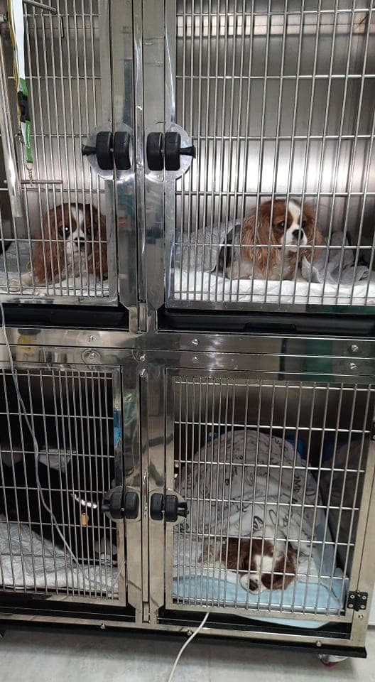
After surgery, patients are kept under observation in the hospital for around 3 hours after waking before happily heading home. This is time during which staff assess whether the animal is breathing properly, whether its body temperature is stable, and whether it has regained enough control over its own body after anaesthesia to function safely outside the hospital. Only once these conditions are met can the patient happily head home, into the owner's care.
This observation period isn't a formality. Right after waking, an animal can be groggy, have a lowered body temperature, and show weakened reflexes – a normal stage of coming round from anaesthesia, but one that takes a trained eye to distinguish from signs that should raise concern, such as breathing difficulty, pale mucous membranes, or excessive bleeding from the wound. Hospital staff have that experience, and it's only after their assessment that a decision is made to discharge the patient.
Follow-Up Checks in the Following Days
Over the following days, the animals are checked by internal medicine veterinarians, who assess the appearance of the wound and the patients' general wellbeing. This follow-up matters just as much as the surgery itself – it's at this stage that any complications are easiest to catch before they have a chance to develop, and to respond to before they become a serious problem.
Preparation and Returning Home
We describe how to prepare your pet for a procedure, and what the first day back home looks like, in detail in a separate article, Before and After a Procedure – covering everything from pre-operative fasting, through what's worth bringing to the clinic, to wound care and the signs that should prompt you to contact us urgently.
Frequently Asked Questions
Does every illness require surgery?
No. The vast majority of conditions you bring to our clinic can be managed with conservative treatment – medication, diet, rehabilitation. We turn to surgery when a problem has a mechanical or structural cause that can't be resolved any other way. The final decision is always made by the treating vet based on complete diagnostics, not on a mere suspicion of illness.
How does inhalation anaesthesia differ from intravenous anaesthesia?
Under inhalation anaesthesia, sleep is maintained with a mixture of gas (isoflurane) and medical oxygen, which the animal breathes, rather than with medication given into a vein. This means the body, and the liver in particular, has less work to do metabolising the anaesthetic. The choice of anaesthesia method is always made by the vet, taking into account the animal's age, breed, weight, and any concurrent illnesses.
Is surgery safe for an older pet?
Age alone isn't a contraindication for surgery – overall health matters more than the number of years. Before any planned operation we recommend additional diagnostics, such as blood tests and sometimes an X-ray or an echocardiogram, which help assess individual risk and, where possible, allow us to choose a safer anaesthesia protocol, such as inhalation anaesthesia.
How long does recovery from surgery take?
That depends on the type of procedure, the animal's age, and its general condition – simple, planned procedures usually come with a shorter recovery than extensive abdominal surgery. The wound itself usually heals fully within a couple of weeks, while a full return to normal activity can take longer, especially after orthopaedic procedures or extensive abdominal surgery. You'll receive a precise healing timeline and detailed post-operative instructions individually from the vet who carried out the procedure.
Is a follow-up visit necessary after surgery?
Yes. In the days following an operation, your pet should be checked by a vet, who will assess how the wound is healing and the patient's general wellbeing. This is a standard part of post-operative care, not an optional extra.
Does every removed tumour need to be sent for histopathology?
We strongly recommend it, even if a growth looks harmless on clinical examination. Histopathology is the only way to establish with certainty what type of growth it was, assess whether the excision margin was sufficient, and determine whether further oncological follow-up is needed. Without this test, both owner and vet are left guessing.
Does surgery always require full anaesthesia?
Not always. Minor procedures are sometimes carried out under sedation, occasionally with local anaesthesia, without the need to fully suppress consciousness or intubate. The scope of the procedure, the animal's temperament, and its state of health determine which option the vet proposes – always assessed individually, case by case. It's worth asking about this at the qualifying visit, so you know what to expect on the day of the procedure.
The decision to operate is never an easy one, but a well-performed procedure – preceded by thorough diagnostics and supported by safe anaesthesia – is often the most effective way to restore an animal's health and comfort. Whether it's a planned castration, the removal of a concerning skin growth, or an urgent intervention after an accident, it helps to have a trusted vet guiding you through the whole process, step by step, from the first consultation, through the procedure itself, to the final follow-up visit. If your vet has recommended surgery for your pet, or if you're unsure whether a particular growth or condition calls for a surgeon's attention, we invite you for a consultation at the Hau-Miau clinic at ul. Siemieńskiego 23 in Warsaw's Ochota district. Book a visit — call +48 22 823 35 63.